
栏目简介:CELL官网 Cell Picture Show(细胞照片秀)栏目(http://www.cell.com/pictureshow),由卡尔蔡司赞助支持,不定期地为大家分享细胞、发育和分子生物学中获得的各种引人注目的照片,让大家欣赏到前沿研究中的美丽图像。
2017 KI Image Awards
Since 2013, Cell Press has partnered with the Koch Institute for Integrative Cancer Research at MIT to feature their annual image award winners in the Cell Picture Show. These images always offer a fascinating glimpse at the worlds opened to us by microscopy and other biomedical imaging techniques, and for that reason, we’re proud to feature the 2017 winners here.
Both the Koch Institute Image Awards and the Cell Picture Show share a similar ethos: Recognizing and disseminating the extraordinary imagery produced through life science research. On March 24, 2017, these winning images were unveiled at the Koch Institute Public Galleries in Cambridge, MA.
We congratulate the 2017 Koch Institute Image Award winners and are excited to continue the collaboration between MIT and Cell Press. This collection represents not only a stunning array of scientific images but also a token of continued collaboration between two neighbors in Cambridge.
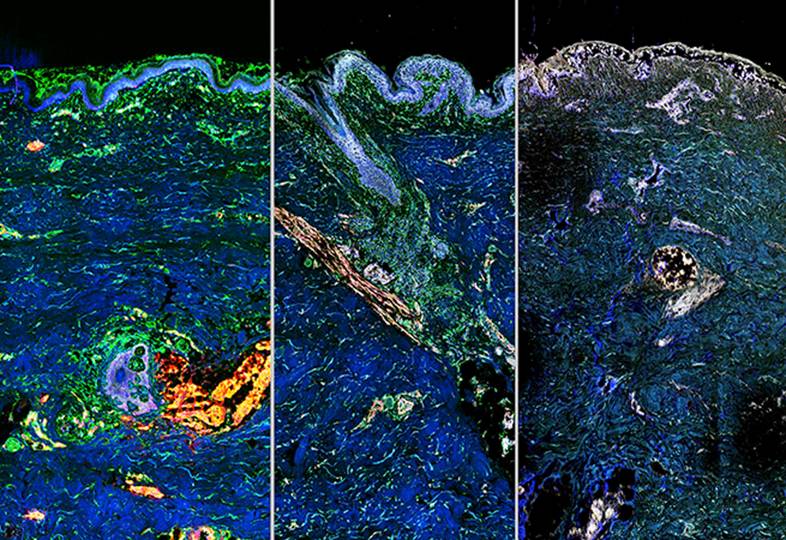
Image: Making Waves: Delivery for Ageless Skin
Carl Schoellhammer, Denitsa Milanova, Humberto Trevino, Cody Cleveland, Jeffrey Wyckoff, Anna Mandinova, Giovanni Traverso, Robert Langer, and George Church
Koch Institute at MIT, Harvard University, and MGH
What if ultrasound (best known for applications in medical imaging and diagnostics) could be used to deliver age-reversing gene therapy to cells?
New technology from the Langer Lab, developed in partnership with Harvard’s Church Lab, drives non-invasive sound waves carrying genetic material through protective layers of skin, seen from top to bottom in these three images. The grey flecks show areas of successful gene transfer to cells, whose genetic clocks have been turned back by the nucleic acids they have received.
View in the Koch Institute Public Galleries
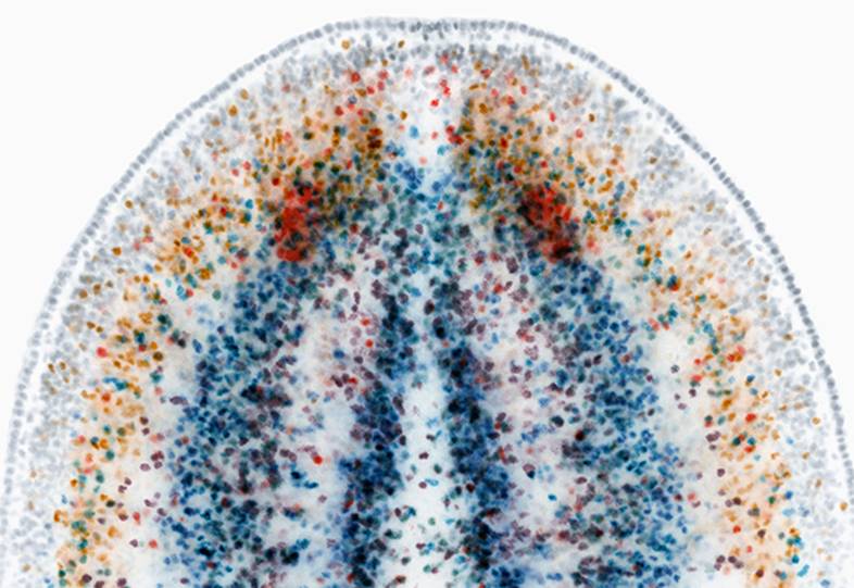
Snap ChAT: A Flatworm Creates a New Profile
Samuel A. LoCascio, Kutay Deniz Atabay, and Peter Reddien
Whitehead Institute
The planarian flatworm shown here possesses a remarkable ability to regenerate complex tissue structures lost to injury, including a brain and eyes. Each color represents a different layer of neurons in the head, revealed by expression of the choline acetyltransferase (ChAT) gene. Not shown in this image are the abundant stem cells that give rise to a new central nervous system as the flatworm regenerates. The Reddien Lab studies planarians to illuminate the cellular and molecular basis of regeneration.
View in the Koch Institute Public Galleries
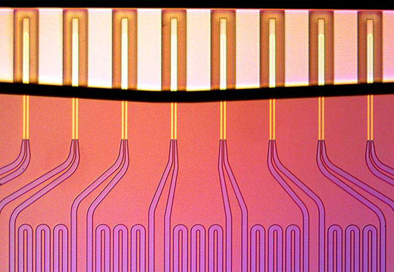
Microfluidics for the Masses: Measuring Cell Growth Rates
Selim Olcum, Nathan Cermak, and Scott Manalis
Koch Institute at MIT
To understand cancer cells’ response to therapy, the Manalis Lab measures how their masses change while exposed to drugs. The fluid-filled channels (bottom) connect tiny mass sensors in the form of hollow diving boards (top) whose vibrations precisely reveal the mass of individual cells passing through them.
As treated cells flow across the array of sensors, each cell is weighed multiple times, thereby revealing the rate at which individual cells change their mass. Researchers are now starting to use tumor cell measurements to predict optimal treatment strategy for individual patients.
View in the Koch Institute Public Galleries
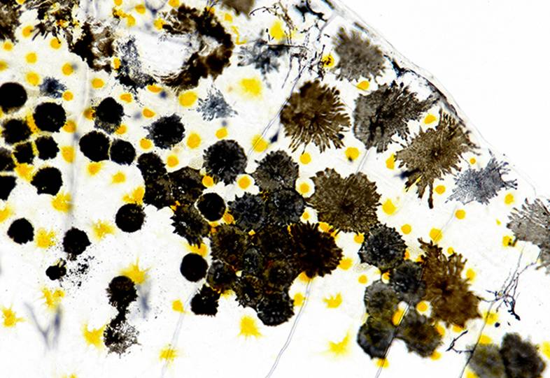
Downstream Dreams: Investigating Melanoma in Zebrafish
Dahlia E. Perez and Jacqueline A. Lees
Koch Institute at MIT
Like a pebble dropped into a pond, a single genetic mutation can trigger a ripple of biological consequences, including cancer. The Lees Lab uses various model systems to track the progression of cancer from origin to disease.
Here, a close-up view of melanocytes in zebrafish gives insight into the organization of these cells in their normal state. Next, biologists will mutate a single gene, a known initiator of uveal melanoma, and study the cells throughout zebrafish development to determine the downstream effects of this single mutational event.
View in the Koch Institute Public Galleries
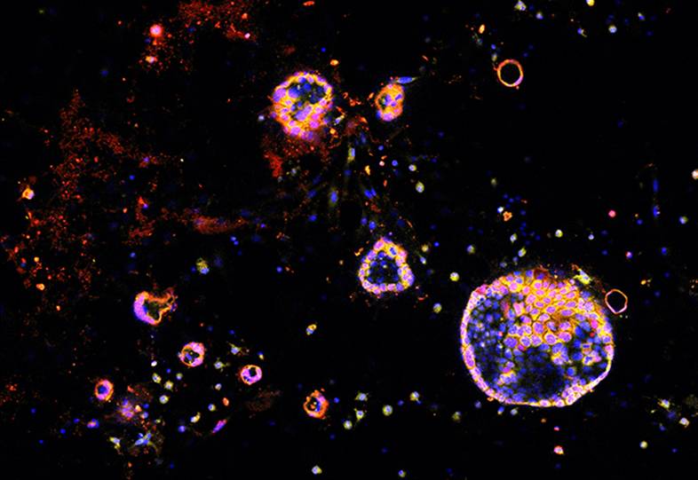
Tiny Trojan Horses: Tumor-Penetrating Nanoparticles Infiltrate Cancer Cells
Liangliang Hao, Srivatsan Raghavan, Emilia Pulver, Jeffrey Wyckoff, and Sangeeta Bhatia
Koch Institute at MIT
You can lead a nanoparticle to tumor cells, but you can’t make them shrink—at least not until the particle gets inside. This image shows biocompatible nanoparticles (yellow) inside clusters of pancreatic cancer cells (pink). The particles’ two-peptide uptake system—one to target the tumor, the second to penetrate it—was specially designed to overcome known difficulties in treating pancreatic cancer, but the Bhatia Lab hopes to expand the use of this modular delivery system for other cancer types as well.
View in the Koch Institute Public Galleries
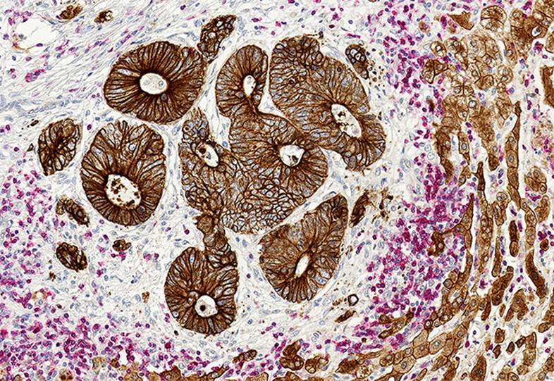
Mind the Gap: Studying the Tumor Extracellular Matrix
Steffen Rickelt and Richard Hynes
Koch Institute at MIT
Although the brightly colored cells in the center of this image catch your eye first, it’s the seemingly neutral tissue around them that the Hynes Lab is studying. Does this extracellular matrix serve as a barrier or a doorway to metastasis?
When cancer cells spread to distant organs, they must navigate a complex network of cells and proteins to get there. This image shows colon cancer metastases (brown clusters) surrounded by liver (light brown) and immune cells (pink). Researchers seek to discover how the surrounding matrix (white/blue) facilitates or limits the interactions between them.
View in the Koch Institute Public Galleries
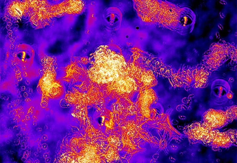
Shape Shifters: Cancer Cells in Motion
Claudia Schafer and Frank Gertler
Koch Institute at MIT
These metastatic lung cancer cells are showing their true nature as they wander around the dish. Images taken ten minutes apart over the course of 16 hours are stacked atop each other to create a composite image. Researchers in the Gertler Lab are studying how different levels of proteins expressed by the cells affect their shape and motion.
Take a look—can you trace the pathways? Are the cells moving slowly or quickly? Do they change shape or stay round? How do they compare to each other?
View in the Koch Institute Public Galleries
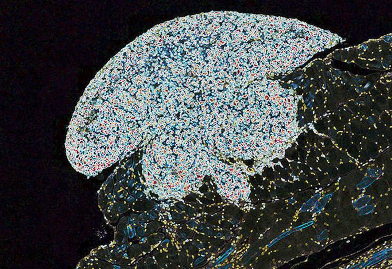
Pushing Boundaries: Ovarian Cancer Hides in Plain Sight
Erik C. Dreaden, Yi Wen Kong, Michael Yaffe, and Paula T. Hammond
Koch Institute at MIT
Persistence is key. Here, an ovarian tumor clings to the abdominal wall, slowly breaking through the tissue boundaries that block its metastatic spread. The tissues here were stained with a molecule that binds to cells that are rapidly growing and multiplying (white). Just as tenacious as these proliferating cells, however, are the researchers who study it. The Yaffe and Hammond Labs are working together to better understand and exploit the genetic weaknesses of these tumors in various disease models, and soon will test their response to experimental treatments, unlocking new avenues for investigation and intervention.
View in the Koch Institute Public Galleries
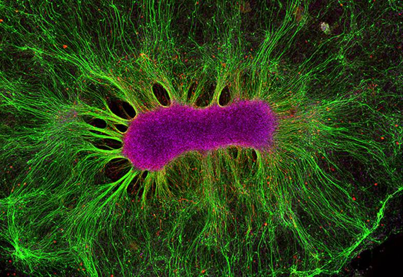
Body of Knowledge: Self-Organizing Brain Cells
Collin Edington, Iris Lee, and Linda Griffith
MIT Department of Biological Engineering and Koch Institute at MIT
Imagine a uniform field of neural stem cells sitting on gel-like matrix. Slowly, they begin to differentiate, grouping and clustering together until they have self-assembled into a mini-organ—a brain!
The neurons (green) and astrocytes (red) seen here are part of the Griffith Lab’s “Human on a Chip” project. Many different mini-organs are linked together in a bioreactor platform, allowing researchers to study the interactions of multiple organs and the crosstalk between them in an in vitro setting and accelerate the development of novel disease treatments.
View in the Koch Institute Public Galleries
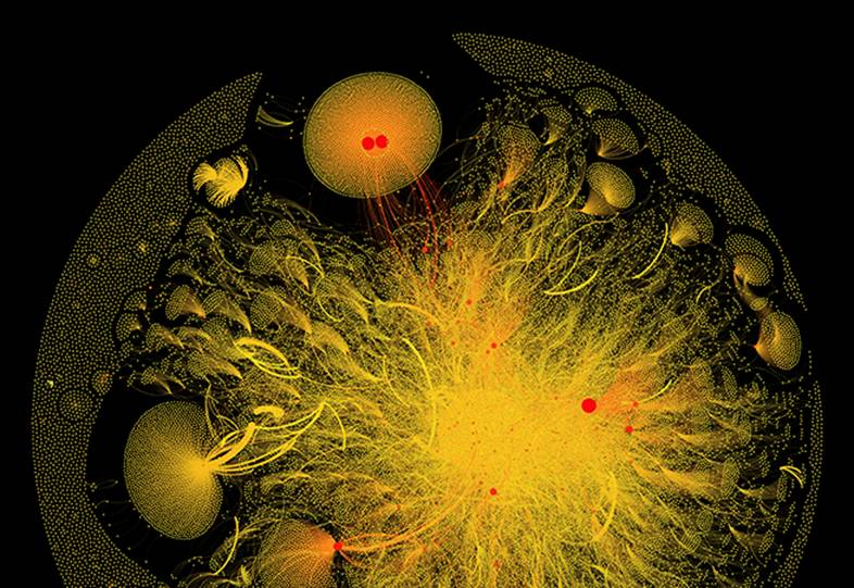
Hashtag No Filter: Visualizing Breast Cancer Conversations
Eric Clarke, Richard Arnett, and Jane Burns
Royal College of Surgeons in Ireland/Wellcome Images
Eight weeks. 92,915 tweets. One hashtag. Users are nodes and the relationships between them are lines.
This image literally visualizes conversations around breast cancer, and the network of connected cancer patients and their loved ones, patient advocates, oncologists, and other healthcare professionals as well as cancer researchers.#breastcancer joins these diverse stakeholders together in one conversation and puts everyone on the same page, erasing societal boundaries to share knowledge and support in real time.
This image appears in the Koch Institute Public Galleries as part of a partnership between the Koch Institute and Wellcome Images.
View in the Koch Institute Public Galleries