
栏目简介:CELL官网 Cell Picture Show(细胞照片秀)栏目(http://www.cell.com/pictureshow),由卡尔蔡司赞助支持,不定期地为大家分享细胞、发育和分子生物学中获得的各种引人注目的照片,让大家欣赏到前沿研究中的美丽图像。
_____________________________________________________________________

http://www.biophysics.org/
Welcome to the Cell Picture Show: Beauty in Biophysics 2017!
After a successful inaugural year, Biophysics Week returns for another round of fascinating microscopic images from the Biophysical Society’s annual image contest. These 10 finalists offer a detailed and comprehensive look into the complex yet beautiful world of biophysics. The winners of the competition were announced at the 61st annual Biophysical Society meeting, which took place in New Orleans on February 11-15. Congratulations to Ryan Littlefield (third place), Eamonn Kennedy (second place), and Giulia Palermo (first place) on being selected.
Biophysics Week is an annual international effort to raise awareness of the field of biophysics, celebrate its accomplishments, and make connections within the biophysics community. Started in 2016, the weeklong celebration returns for the second time from March 6-10, 2017.
欢迎来到细胞图片展示:美容生物物理2017!
在成功的开始一年后,生物物理周再次从生物物理学会的年度形象大赛中获取另一轮迷人的显微镜图像。这10个入围者对复杂但美丽的生物物理世界提供了详细和全面的看法。比赛的获胜者在第61 届年度生物物理学会会议上宣布,该会议于2月11-15日在新奥尔良举行。恭喜Ryan Littlefield(第三名),Eamonn Kennedy(第二名)和Giulia Palermo(第一名)被选中。
生物物理周是一项年度国际努力,以提高对生物物理学领域的认识,庆祝其成就,并在生物物理学界内建立联系。从2016年开始,这一周的庆祝活动将于2017年3月6日至10日举行第二次。
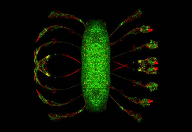
Tensed to Sense
Anthony Fan
University of Illinois
Neurons (green) and cytoskeletons (red) of a developing fruit fly embryo imaged under a confocal microscope. When buckled (left)—i.e. by a compressive load—neurons would straighten (right) in a matter of a few minutes. This is achieved by an axial contraction generated and maintained by various cytoskeletal proteins.
感觉到
伊利诺伊州安东尼·范大学
在共焦显微镜下成像的发育中的果蝇胚胎的神经元(绿色)和细胞骨架(红色)当压曲(左) - 通过压缩负载 - 神经元将在几分钟内伸直这是通过由各种细胞骨架蛋白产生和维持的轴向收缩实现的。
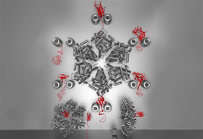
Atomistic Simulations of Mechanical Protein Remodeling by ClpATPase Nanomachines
Abdolreza Javidialesaadi
University of Cincinnati
An implicit solvent model of ClpYΔI-mediated unfolding and translocation of the immunoglobulin protein domain Titin I27. Cyclical opening and closing of the ClpYΔI ring are modeled through sequential conformational changes of ClpYΔI subunits. Simulations were performed using CHARMM. VMD and Blender were used for rendering.
机械蛋白重塑的原子模拟由ClpATPase纳米机械
辛辛那提大学
Abdolreza Javidialesaadi
ClpYΔI介导的免疫球蛋白结构域Titin I27的解折叠和易位的隐性溶剂模型。通过ClpYΔI亚基的顺序构象变化来模拟ClpYΔI环的循环开放和闭合。使用CHARMM进行模拟。VMD 和Blender用于渲染。
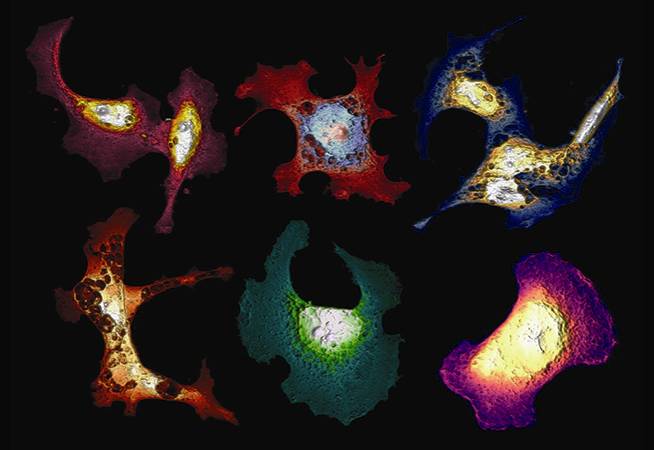
A Morphological Examination of Programmed Cell Death
Eamonn Kennedy, Rasoul Al-Majmaie, and James Rice
University of Notre Dame and University College Dublin
A composite of six atomic force microscopy (AFM) topographs of SW480 colon cancer cells. Programmed cell death (apoptosis) has been induced by laser irradiation after uptake of a gold nanoparticle-conjugated photo-sensitizer, resulting in common features of apoptosis such as pore formation and changes to membrane roughness (evident in the height-to-color images). Given that these features are both three-dimensional and sub-micron in scale, AFM is an ideal method for their detection. Congratulations to Eamonn Kennedy for winning second place in the Biophysical Society’s annual image contest.
程序性细胞死亡的形态学检查
Eamonn Kennedy,Rasoul Al-Majmaie和James Rice
大学圣母大学和都柏林大学
SW480结肠癌细胞的六原子力显微镜(AFM)形状的复合物。在摄取金纳米颗粒共轭的光敏剂后,通过激光照射诱导程序性细胞死亡(凋亡),导致细胞凋亡的共同特征,例如孔形成和膜粗糙度的变化(在高度至彩色图像中是明显的)。考虑到这些特征在尺度上都是三维和亚微米,AFM是它们的检测的理想方法。恭喜Eamonn Kennedy在生物物理学会年度图片大赛中获得第二名。
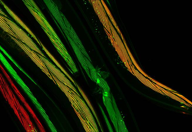
Fluorescent Muscles in Living C. elegans
Ryan Littlefield
University of South Alabama
Transgenic Caenorhabditis elegans expressing GFP-Tropomodulin (green) and mCherry-myosin (headless mutant of minor isoform A; red). Prominent striations within body wall muscle show normal sarcomeric organization and that overlap between the thin and thick filaments is preserved when the myosin A isoform is absent. Images are the maximum intensity projection of overlapping fields acquired by spinning disk confocal microscopy. Congratulations to Ryan Littlefield for winning second place in the Biophysical Society’s annual image contest.
荧光肌在生活线虫
南阿拉巴马州Ryan Littlefield 大学
转基因秀丽隐杆线虫 表达GFP-Tropomodulin(绿色)和mCherry肌球蛋白(未成年人亚型A的无头突变体;红色)。体壁肌肉中突出的条纹显示正常的肌节组织,并且当肌球蛋白A同种型不存在时保持细和粗丝之间的重叠。图像是通过旋转盘共聚焦显微镜获得的重叠场的最大强度投影。恭喜Ryan Littlefield在生物物理学会的年度形态大赛中获得第二名。
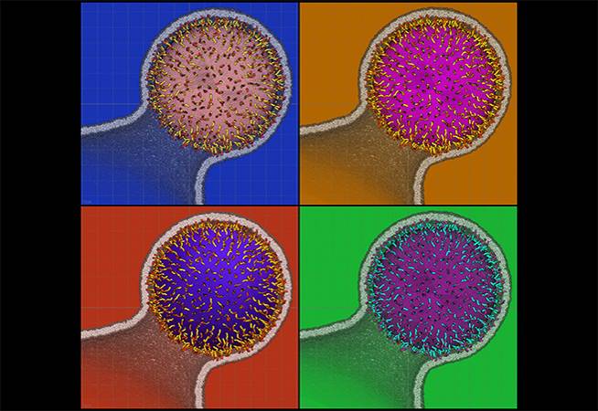
Influenza Budding Prints
Jesper Madsen, John M. A. Grime, and Gregory A. Voth
University of Chicago
Snapshots from a molecular simulation of budding where the membrane envelope has a constricted neck. A spherical polymer brush “fuzzball” guides the membrane shape by an attractive interaction; a minimalist method for representing the effects of the protein coat in influenza virus. Inspired by Andy Warhol and Roy Lichtenstein.
流感发芽印刷品
耶斯帕·马德森,约翰·马格里姆和格雷戈里·沃思
芝加哥大学
来自分子模拟的芽生的快照,其中膜包膜具有收缩的颈部。球形聚合物刷“模糊球”通过有吸引力的相互作用引导膜形状; 一种用于表示流感病毒中蛋白质外壳的影响最低限度的方法。灵感来自安迪·沃霍尔和罗伊·利希滕斯坦。
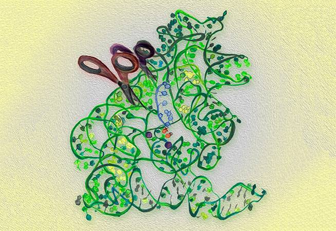
Handmade Painting of Group II Intron Robozyme
Giulia Palermo, Lorenzo Casalino, Giulia Palermo, Ursula Rothlisberger, and Alessandra Magistrato
University of California, San Diego
Handmade picture of Group II intron, considered the bacterial ancestors of the spliceosome, which is a key biological machinery that performs the conversion of pre-mature mRNA into mature mRNA in humans. Two shares are used to highlight the splicing mechanism. Congratulations to Giulia Palermo for winning first place in the Biophysical Society’s annual image contest.
第二组内含子Robozyme的手工绘画
Giulia Palermo,Lorenzo Casalino,Giulia Palermo,Ursula Rothlisberger和Alessandra Magistrato
加利福尼亚大学,圣地亚哥
II组内含子的手工图片,被认为是剪接体的细菌祖先,其是在人类中执行成熟mRNA的关键生物机械。两个份额用于突出拼接机制。祝贺Giulia Palermo在生物物理学会的年度形象大赛中获得第一名。
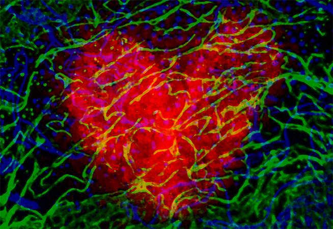
Enchain My Art
Patrice Rassam
University of Oxford
Escherichia coli produce protein antibiotics in order to kill other bacteria competing for life. To better understand these potential therapeutics, we have developed fluorescent labels and use confocal multicolor microscopy (Zeiss LSM780). Here, we have managed to identify how a couple of bacteria could resist antibiotic treatment by hiding in a biofilm.
Enchain我的艺术
牛津大学Patrice Rassam 大学
大肠杆菌产生蛋白质抗生素以杀死竞争生命的其他细菌。为了更好地了解这些潜在的治疗,我们已经开发了荧光标记和使用共焦多色显微镜(蔡司LSM780)。在这里,我们设法确定如何一对夫妇的细菌可以通过隐藏在生物膜抵抗抗生素治疗。
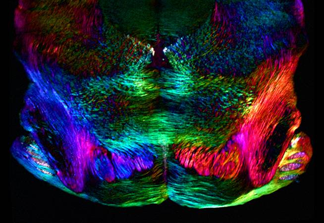
Polarization Polychromatic Image of Mouse Midbrain Slice
Michael Shribak
Marine Biological Laboratory
A mouse midbrain section image obtained with new polychromatic polscope, which allows us to directly see the colored polarization image, with brightness corresponding to retardance and color corresponding to the slow axis azimuth. The image shows a coronal view of 20 µm-thick section of the brain, which has been sliced down a vertical axis.
The picture was taken in white light without any filter. No staining was applied to the transparent specimen. Nerve cells appear in the real colors that reveal orientation of the molecules. We used an inverted light microscope (Olympus IX81) equipped with an objective lens (UPlanFL 4 x/0.13, zoom lens 0.5 x and 100 W halogen lamp U-LH100-3-5). The images were captured with an Olympus color CCD camera DP73. The image size is 2.9 mm x 2.1 mm.
鼠标中脑切片的偏振多色图像
迈克尔Shribak
海洋生物实验室
用新的多色波导获得的小鼠中脑部分图像,其允许我们直接看到彩色偏振图像,具有对应于慢轴方位角的延迟和颜色的亮度。该图显示了脑的厚度为20μm的部分其已经沿垂直轴切下。
图片是在没有任何过滤器的白色光。没有对透明样品施加染色。神经细胞出现在显示分子取向的真实颜色中。我们使用配备有物镜(UPlanFL 4x / 0.13,变焦透镜0.5x和100W卤素灯U-LH100-3-5)的倒置光学显微镜(Olympus IX81)。使用Olympus彩色CCD照相机DP73捕获图像图像大小为2.9mm×2.1mm。
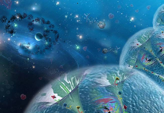
MLKL forms Cation Channels
Bingqing Xia
Shanghai Institute of Materia Medica Chinese Academy of Sciences
MLKL, the necroptosis executioner, forms a new type of cation channels that are permeable to Mg2+, Na+, and K+. The channel activity is positively correlated to its capability to induce cell death. Thus, MLKL channels connect the programmed necrosis with extracellular death signals just like the wormhole connects two dimensional spaces. This picture is featured on the cover image of Cell Research, volume 26, number 5.
MLKL形成阳离子通道
冰清夏
本草中国中科院上海研究所
MLKL,坏死死亡执行者,形成一种新型的阳离子通道,可渗透的Mg2 +,钠+和ķ +。通道活性与其诱导细胞死亡的能力正相关。因此,MLKL通道将程序性坏死与细胞外死亡信号连接起来,就像虫洞连接二维空间一样。这张图片在Cell Research,第26卷,第5期的封面图片上。
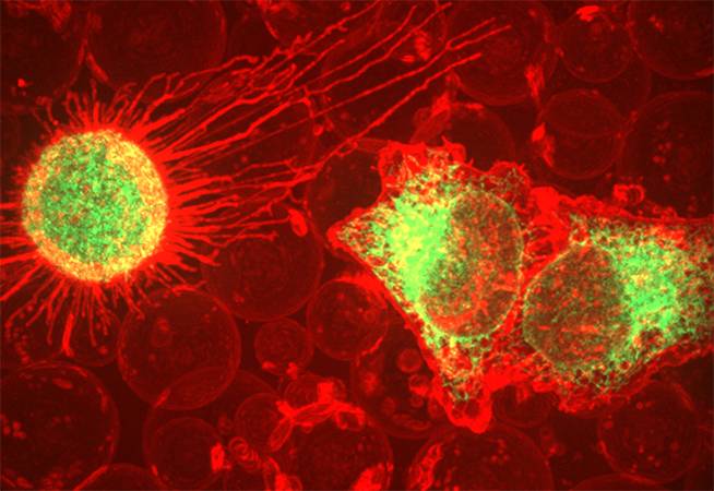
Cell-Derived Plasma Membrane Vesicles Interacting with Target Cells
Chi Zhao, David J. Busch, Connor P. Vershel, and Jeanne C. Stachowiak
The University of Texas
A 3D reconstruction taken with a spinning disc confocal microscope. The image depicts the interaction between cell-derived plasma membrane vesicles and target cells expressing GFP-tagged receptors.
细胞衍生的等离子膜囊泡与目标细胞相互作用
Chi Zhao,David J.Busch,Connor P.Verhel和Jeanne C.Stachowiak得克萨斯
大学
用转移的圆盘共焦显微镜采样的3D重建。该图像描述了细胞衍生的质膜囊泡和表达GFP标记的受体的靶细胞之间的相互作用。